What is a vascular ultrasound?
Vascular ultrasounds are noninvasive imaging tests that use high-frequency sound waves to create images, also known as sonograms, of your arteries and veins. Also called vascular sonography, it reveals the movement and structure of your internal organs and how blood flows within your vessels.
Your vascular system is made up of an intricate system of blood vessels that bring blood to and from vital organs, including your heart and brain. Vascular ultrasounds can help you and your physician identify vascular diseases that increase your risk for heart attacks, strokes and other serious conditions, allowing you to be proactive about your health.
When is a vascular ultrasound performed?
Your provider may recommend vascular ultrasound if you have symptoms of the following conditions:
- Atherosclerosis, when the arteries become narrowed and hardened due to the buildup of plaque
- Blood clots
- Carotid artery disease
- Carotid artery stenosis, the narrowing of one or both carotid arteries
- Deep vein thrombosis or leg swelling
- Abdominal aortic aneurysms
- Peripheral artery disease (PAD)
- Peripheral vascular disease (PVD)
- Varicose veins
You may also need ultrasound imaging if your doctor wants to:
- Assess whether a treatment or procedure is treating your condition effectively
- Help determine whether you may benefit from a treatment such as angioplasty
- Monitor blood flow to organs and tissues
Types of vascular ultrasound
Depending on your symptoms, your doctor may recommend one or more of the following vascular ultrasounds:
Aortic ultrasound
Carotid ultrasound
Venous ultrasound
Ankle brachial index (ABI)
What to expect with vascular ultrasound
Before a vascular ultrasound
Vascular ultrasounds don’t require much special preparation. You may receive instructions specific to your test, but in general, you may need to:
- Avoid eating or drinking for a few hours before your test.
- Take any medications as prescribed.
- Wear a two-piece outfit (no dresses, jumpsuits or overalls).
During a vascular ultrasound
Before your exam begins, a sonographer will place gel on the area being examined. Then, they’ll use a device called a transducer that sends sound waves through your skin. These sound waves return to the transducer, and a computer translates them into images that give your provider a clear view of issues in your aorta, carotid arteries and veins.
Aorta and venous ultrasounds take about one hour. A carotid ultrasound takes about 30 minutes.
After a vascular ultrasound
After the exam, the technologist will wipe away any residual gel. You can return to work or other activities immediately after the ultrasound.The results
After your ultrasound, your sonographer will send your images to your referring physician, who will review your results and compare them with any previous tests you’ve had.
Based on the results, your doctor may recommend other tests to confirm a diagnosis. If the ultrasound leads to a diagnosis or reveals a problem, you and your physician can work together on a treatment plan.
Find a location near you
Vascular ultrasounds are available at Baylor Scott & White locations across Central and North Texas. If your ultrasound leads to a diagnosis, you can access comprehensive vascular care at a location convenient to you.

Baylor Scott & White All Saints Medical Center - Fort Worth
1400 8th Ave , Fort Worth, TX, 76104
Baylor Scott & White Clinic - Temple
2401 S 31st St , Temple, TX, 76508- Monday: 8:00 am - 5:00 pm
- Tuesday: 8:00 am - 5:00 pm
- Wednesday: 8:00 am - 5:00 pm
- Thursday: 8:00 am - 5:00 pm
- Friday: 8:00 am - 5:00 pm
Baylor Scott & White Heart and Vascular Hospital - Dallas
621 N Hall St , Dallas, TX, 75226
Baylor Scott & White Heart and Vascular Hospital - Fort Worth
1400 8th Ave Bldg A, 6th Floor, Fort Worth, TX, 76104
Baylor Scott & White Medical Center - Centennial
12505 Lebanon Rd , Frisco, TX, 75035
Baylor Scott & White Medical Center - Frisco
5601 Warren Pkwy , Frisco, TX, 75034
Baylor Scott & White Medical Center - Grapevine
1650 W College St , Grapevine, TX, 76051
Baylor Scott & White Medical Center - Hillcrest
100 Hillcrest Medical Blvd , Waco, TX, 76712
Baylor Scott & White Medical Center - Irving
1901 N MacArthur Blvd , Irving, TX, 75061
Baylor Scott & White Medical Center - Lake Pointe
6800 Scenic Dr , Rowlett, TX, 75088
Baylor Scott & White Medical Center - Lakeway
100 Medical Pkwy , Lakeway, TX, 78738
Baylor Scott & White Medical Center - Marble Falls
810 W State Hwy 71 , Marble Falls, TX, 78654
Baylor Scott & White Medical Center - McKinney
5252 W University Dr Highway 380 at Lake Forest Drive, McKinney, TX, 75071
Baylor Scott & White Medical Center - Plano
4700 Alliance Blvd , Plano, TX, 75093
Baylor Scott & White Medical Center - Round Rock
300 University Blvd , Round Rock, TX, 78665
Baylor Scott & White Medical Center - Sunnyvale
231 S Collins Rd , Sunnyvale, TX, 75182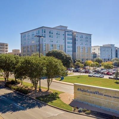
Baylor Scott & White Medical Center - Temple
2401 S 31st St , Temple, TX, 76508
Baylor Scott & White Medical Center - Waxahachie
2400 N Interstate 35E , Waxahachie, TX, 75165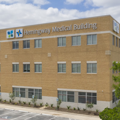
Baylor Scott & White Specialty Clinic - Killeen Hemingway
2405 S Clear Creek Rd , Killeen, TX, 76549- Monday: 8:00 am - 5:00 pm
- Tuesday: 8:00 am - 5:00 pm
- Wednesday: 8:00 am - 5:00 pm
- Thursday: 8:00 am - 5:00 pm
- Friday: 8:00 am - 5:00 pm
Baylor Scott & White Specialty Clinic - Lakeway
200 Medical Pkwy , Lakeway, TX, 78738- Monday: 8:00 am - 5:00 pm
- Tuesday: 8:00 am - 5:00 pm
- Wednesday: 8:00 am - 5:00 pm
- Thursday: 8:00 am - 5:00 pm
- Friday: 8:00 am - 5:00 pm
Baylor Scott & White The Heart Hospital - Denton
2801 S Mayhill Rd , Denton, TX, 76208
Baylor Scott & White The Heart Hospital - McKinney
5268 W University Dr , McKinney, TX, 75071
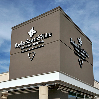
Baylor Scott & White The Heart Hospital - Plano
1100 Allied Dr , Plano, TX, 75093
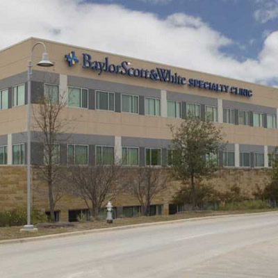
Baylor Scott & White Vascular and Vein Clinic - Austin
2217 Park Bend Dr Ste 230, Austin, TX, 78758- Monday: 8:00 am - 5:00 pm
- Tuesday: 8:00 am - 5:00 pm
- Wednesday: 8:00 am - 5:00 pm
- Thursday: 8:00 am - 5:00 pm
- Friday: 8:00 am - 5:00 pm
Baylor Scott & White Vascular Surgery Specialists - Fort Worth
1250 8th Ave Ste 200, Fort Worth, TX, 76104- Monday: 8:30 am - 4:30 pm
- Tuesday: 8:30 am - 4:30 pm
- Wednesday: 8:30 am - 4:30 pm
- Thursday: 8:30 am - 4:30 pm
- Friday: 8:30 am - 4:30 pm
Baylor Scott & White Vascular Surgery Specialists - Grapevine
2020 W State Hwy 114 Ste 200, Grapevine, TX, 76051
Baylor Scott & White Vascular Surgery Specialists - Mansfield
1776 N US 287 Ste 220, Mansfield, TX, 76063