Detect and monitor heart disease with cardiac imaging
Cardiac imaging captures images of your heart. Your provider may order cardiac imaging to evaluate your risk for cardiovascular conditions, investigate the cause of your symptoms or confirm a diagnosis. You may also need cardiac imaging to determine if a medication or other heart disease treatment is working properly.
When is cardiac imaging performed?
Your cardiologist can use cardiac imaging to detect many heart and vascular conditions. A few of them include:
Cardiac computed tomography (CT)
A cardiac CT scan of the heart takes multiple X-ray images to create a detailed, 3D picture of your heart muscle and blood vessels. In some cases, your doctor may use a contrast dye to get a better view of your blood vessels.
Contrast dyes often contain iodine, which can cause a reaction in people with an iodine allergy. Let your imaging team know beforehand if you know you have an iodine allergy. If you do, they can use steroids and antihistamines to prevent an allergic reaction.
Cardiac CT scans can show:
- Blockages in your arteries, which may contribute to aneurysms and heart attacks
- Calcium deposits in your arteries, which is used to determine your calcium score, a test that can help determine your risk for heart attack or other heart events
- How well blood flows through the heart, which helps determine if you have heart valve issues
- How well your heart pumps blood, which can help your doctor understand your risk for heart failure
What to expect during cardiac CT
- Cardiac CT scanners are large, doughnut-shaped devices. You lie on a padded table that slides through the scanner’s opening, and the machine takes X-ray images of your heart.
- You will need to lie very still, and the radiologic technologist may ask you to hold your breath to minimize body movement while the images are taken. The technologist will monitor you during the entire exam through a window and communicate with you through an intercom if you need help or feel uncomfortable.
- The length of the exams can vary but plan on the test-taking up to an hour. Tests that involve dyes may take longer.
- After the test, a radiologist will read your images and send a report to your physician, who will discuss the results with you and plan the next steps.
Cardiac positron emission tomography (PET) scans
Sometimes called nuclear imaging, cardiac PET scans combine imaging with a special radioactive substance that helps doctors more easily identify heart problems, such as:
- Damage a heart attack may have caused
- Issues with how blood flows through your heart, which can help them diagnose coronary artery disease
- Whether you would benefit from a specific treatment, such as angioplasty and stenting or coronary artery bypass grafting
Cardiac PET scans may also be used during a type of heart stress test called a pharmacologic stress test. This test helps doctors understand how well your heart works while it’s under stress. Before the test begins, you receive medication that makes your heart beat faster. Images of your heart are then taken with a PET scanner.
Before a heart PET scan, you receive a small amount of radioactive tracer called a radionuclide. The PET scanner detects the tracer and creates detailed 3D images.
The radioactive tracer poses very little risk to most people. Your body eliminates it through urine and bowel movements. However, women who are pregnant, nursing or who think they may be pregnant should not receive cardiac PET scans.
What to expect during a cardiac PET scan
The tests begin with you having an IV placed in a vein in your arm so you can receive the radioactive tracers. You’ll also have a blood pressure cuff placed on one arm and electrodes placed on your chest so your imaging team can monitor your blood pressure and take an electrocardiogram during the test. If you’re having a pharmacologic stress test, you will also receive medication to dilate your arteries.
When ready for imaging, you will be asked to lie on an exam table with your arms above your head. The PET scanner will take images from different angles. Plan for the test to last at least 90 minutes.
After the test, the imaging team will read your images and send a report to your physician, who will discuss the results with you and plan the next steps.
Additional types of cardiac imaging
In addition to cardiac CT and PET scans, your provider may use the following imaging tests:
- Cardiac angiography, an interventional cardiology procedure in which a small tube is inserted into a vein and guided to your heart, where X-ray images and dyes are used to identify blockages
- Echocardiogram, which uses sound waves to produce images of your heart muscle’s size and shape
- X-rays, which show the size, location and shape of your heart and blood vessels
Your provider may recommend one or multiple tests, based on your symptoms and the information they hope to learn from your images.
Preparing for cardiac imaging
Each cardiac imaging test is slightly different. Before your appointment, you will receive instructions specific to the test you are having about how to prepare, what to expect and how to receive your results. These instructions will be specific to the test you are having done.
Not all imaging exams require special preparation, but in general, you can use these tips to prepare for your exam:
- Bring your medications or a list of medicines you take if your care team has asked you to.
- Follow the instructions you receive about eating, drinking and taking medications before your exam.
- Wear loose, comfortable clothing. Short sleeves are a good option if your test requires an IV or involves blood pressure cuffs. Avoid boots, jumpsuits, overalls and dresses.
Find a cardiac imaging location near you
Cardiac imaging is available at many Baylor Scott & White locations. No matter where you go, you will have access to advanced imaging technologies and experienced technologists and radiologists dedicated to helping you get accurate results.

Baylor Scott & White Advanced Heart Failure Clinic - Abilene
1219 E South 11th St Ste B2, Abilene, TX, 79602
Baylor Scott & White Advanced Heart Failure Clinic - Amarillo
1901 Medi Park Dr Ste 2051, Amarillo, TX, 79106
Baylor Scott & White Advanced Heart Failure Clinic - Dallas
3410 Worth St Ste 250, Dallas, TX, 75246- Monday: 8:00 am - 4:30 pm
- Tuesday: 8:00 am - 4:30 pm
- Wednesday: 8:00 am - 4:30 pm
- Thursday: 8:00 am - 4:30 pm
- Friday: 8:00 am - 4:30 pm

Baylor Scott & White Advanced Heart Failure Clinic - Fort Worth
1400 8th Ave Ste 105, Fort Worth, TX, 76104- Monday: 8:00 am - 4:30 pm
- Tuesday: 8:00 am - 4:30 pm
- Wednesday: 8:00 am - 4:30 pm
- Thursday: 8:00 am - 4:30 pm
- Friday: 8:00 am - 4:30 pm

Baylor Scott & White Advanced Heart Failure Clinic - Longview
906 Judson Rd , Longview, TX, 75601
Baylor Scott & White Advanced Heart Failure Clinic - Lubbock
3711 22nd St Ste B, Lubbock, TX, 79410
Baylor Scott & White Advanced Heart Failure Clinic - Midland/Odessa
420 E 6th St Ste 102, Odessa, TX, 79761
Baylor Scott & White Advanced Heart Failure Clinic - Tyler
1321 S Beckham Ave , Tyler, TX, 75701
Baylor Scott & White Advanced Heart Failure Specialists - Fort Worth
1250 8th Ave Ste 200, Fort Worth, TX, 76104- Monday: 8:00 am - 5:00 pm
- Tuesday: 8:00 am - 5:00 pm
- Wednesday: 8:00 am - 5:00 pm
- Thursday: 8:00 am - 5:00 pm
- Friday: 8:00 am - 5:00 pm

Baylor Scott & White All Saints Medical Center - Fort Worth
1400 8th Ave , Fort Worth, TX, 76104
Baylor Scott & White Arrhythmia Management - Denton
3333 Colorado Blvd , Denton, TX, 76210- Monday: 8:00 am - 5:00 pm
- Tuesday: 8:00 am - 5:00 pm
- Wednesday: 8:00 am - 5:00 pm
- Thursday: 8:00 am - 5:00 pm
- Friday: 8:00 am - 5:00 pm

Baylor Scott & White Arrhythmia Management - Garland
7217 Telecom Pkwy Ste 205, Garland, TX, 75044- Monday: 8:00 am - 5:00 pm
- Tuesday: 8:00 am - 5:00 pm
- Wednesday: 8:00 am - 5:00 pm
- Thursday: 8:00 am - 5:00 pm
- Friday: 8:00 am - 5:00 pm

Baylor Scott & White Arrhythmia Management - Grapevine
2020 W State Hwy 114 Ste 320, Grapevine, TX, 76051
Baylor Scott & White Arrhythmia Management - McKinney
5236 W University Dr POB I, Ste 4900, McKinney, TX, 75071- Monday: 8:00 am - 5:00 pm
- Tuesday: 8:00 am - 5:00 pm
- Wednesday: 8:00 am - 5:00 pm
- Thursday: 8:00 am - 5:00 pm
- Friday: 8:00 am - 5:00 pm

Baylor Scott & White Arrhythmia Management - Plano
1820 Preston Park Blvd Ste 1450, Plano, TX, 75093- Monday: 8:00 am - 5:00 pm
- Tuesday: 8:00 am - 5:00 pm
- Wednesday: 8:00 am - 5:00 pm
- Thursday: 8:00 am - 5:00 pm
- Friday: 8:00 am - 5:00 pm

Baylor Scott & White Arrhythmia Management - Prosper
111 S Preston Rd Ste 10, Prosper, TX, 75078- Monday: 8:00 am - 5:00 pm
- Tuesday: 8:00 am - 5:00 pm
- Wednesday: 8:00 am - 5:00 pm
- Thursday: 8:00 am - 5:00 pm
- Friday: 8:00 am - 5:00 pm

Baylor Scott & White Arrhythmia Management - Rockwall
1005 W Ralph Hall Pkwy Ste 225, Rockwall, TX, 75032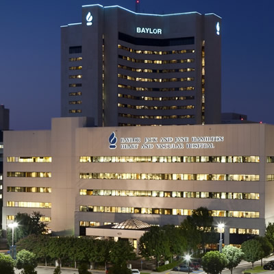
Baylor Scott & White Cardiac Surgery - Dallas
621 N Hall St Ste 120, Dallas, TX, 75226- Monday: 8:30 am - 4:30 pm
- Tuesday: 8:30 am - 4:30 pm
- Wednesday: 8:30 am - 4:30 pm
- Thursday: 8:30 am - 4:30 pm
- Friday: 8:30 am - 4:30 pm

Baylor Scott & White Cardiology Consultants of Texas - Dallas
621 N Hall St Ste 500, Dallas, TX, 75226- Monday: 8:00 am - 5:00 pm
- Tuesday: 8:00 am - 5:00 pm
- Wednesday: 8:00 am - 5:00 pm
- Thursday: 8:00 am - 5:00 pm
- Friday: 8:00 am - 5:00 pm
- Monday: 8:30 am - 4:30 pm
- Tuesday: 8:30 am - 4:30 pm
- Wednesday: 8:30 am - 4:30 pm
- Thursday: 8:30 am - 4:30 pm

Baylor Scott & White Cardiology Consultants of Texas - Greenville
4400 Interstate 30 W Ste 300, Greenville, TX, 75402- Monday: 8:30 am - 5:00 pm
- Tuesday: 8:30 am - 5:00 pm
- Wednesday: 8:30 am - 5:00 pm
- Thursday: 8:30 am - 5:00 pm
- Friday: 8:30 am - 5:00 pm
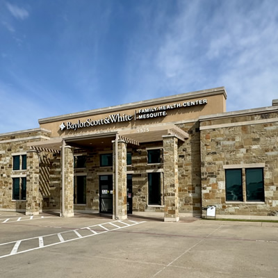
Baylor Scott & White Cardiology Consultants of Texas - Mesquite
1575 Interstate 30 , Mesquite, TX, 75150- Monday: 8:30 am - 5:00 pm
- Tuesday: 8:30 am - 5:00 pm
- Wednesday: 8:30 am - 5:00 pm
- Thursday: 8:30 am - 5:00 pm
- Friday: 8:30 am - 5:00 pm

Baylor Scott & White Cardiology Consultants of Texas - Midway
4431 E US Hwy 287 , Midlothian, TX, 76065- Monday: 8:00 am - 5:00 pm
- Tuesday: 8:00 am - 5:00 pm
- Wednesday: 8:00 am - 5:00 pm
- Thursday: 8:00 am - 5:00 pm
- Friday: 8:00 am - 5:00 pm

Baylor Scott & White Cardiology Consultants of Texas - Park Cities
9101 N Central Expy Ste 300C, Dallas, TX, 75231- Monday: 8:30 am - 5:00 pm
- Tuesday: 8:30 am - 5:00 pm
- Wednesday: 8:30 am - 5:00 pm
- Thursday: 8:30 am - 5:00 pm
- Friday: 8:30 am - 5:00 pm

Baylor Scott & White Cardiology Consultants of Texas - Red Oak
301 E Ovilla Rd Ste 100, Red Oak, TX, 75154- Monday: 8:00 am - 5:00 pm
- Tuesday: 8:00 am - 5:00 pm
- Wednesday: 8:00 am - 5:00 pm
- Thursday: 8:00 am - 5:00 pm
- Friday: 8:00 am - 5:00 pm
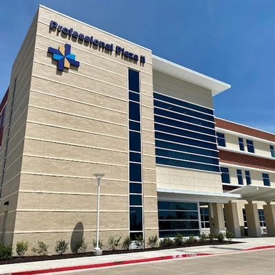
Baylor Scott & White Cardiology Consultants of Texas - Waxahachie
2360 N Interstate 35E Ste 110, Waxahachie, TX, 75165- Monday: 8:00 am - 5:00 pm
- Tuesday: 8:00 am - 5:00 pm
- Wednesday: 8:00 am - 5:00 pm
- Thursday: 8:00 am - 5:00 pm
- Friday: 8:00 am - 5:00 pm

Baylor Scott & White Cardiothoracic Surgery - Irving
1110 Cottonwood Ln Ste 215, Irving, TX, 75038- Monday: 8:30 am - 4:30 pm
- Tuesday: 8:30 am - 4:30 pm
- Wednesday: 8:30 am - 4:30 pm
- Thursday: 8:30 am - 4:30 pm
- Friday: 8:30 am - 4:30 pm

Baylor Scott & White Cardiovascular Associates - Fort Worth
1307 8th Ave Ste 406, Fort Worth, TX, 76104- Monday: 8:00 am - 4:30 pm
- Tuesday: 8:00 am - 4:30 pm
- Wednesday: 8:00 am - 4:30 pm
- Thursday: 8:00 am - 4:30 pm
- Friday: 8:00 am - 4:30 pm

Baylor Scott & White Cardiovascular Consultants - Denton
3333 Colorado Blvd , Denton, TX, 76210- Monday: 8:30 am - 5:00 pm
- Tuesday: 8:30 am - 5:00 pm
- Wednesday: 8:30 am - 5:00 pm
- Thursday: 8:30 am - 5:00 pm
- Friday: 8:30 am - 5:00 pm

Baylor Scott & White Cardiovascular Consultants - Flower Mound
4421 Long Prairie Rd Ste 200, Flower Mound, TX, 75028- Monday: 8:30 am - 5:00 pm
- Tuesday: 8:30 am - 5:00 pm
- Wednesday: 8:30 am - 5:00 pm
- Thursday: 8:30 am - 5:00 pm
- Friday: 8:30 am - 5:00 pm

Baylor Scott & White Cardiovascular Consultants - Grapevine
2020 W State Hwy 114 Ste 200, Grapevine, TX, 76051- Monday: 8:00 am - 5:00 pm
- Tuesday: 8:00 am - 5:00 pm
- Wednesday: 8:00 am - 5:00 pm
- Thursday: 8:00 am - 5:00 pm
- Friday: 8:00 am - 4:00 pm

Baylor Scott & White Cardiovascular Consultants - Keller
620 S Main St Ste 240, Keller, TX, 76248- Monday: 8:00 am - 5:00 pm
- Tuesday: 8:00 am - 5:00 pm
- Wednesday: 8:00 am - 5:00 pm
- Thursday: 8:00 am - 5:00 pm
- Friday: 8:00 am - 4:00 pm

Baylor Scott & White Cardiovascular Consultants - Plano
6000 W Spring Creek Pkwy Ste 220, Plano, TX, 75024- Monday: 8:30 am - 5:00 pm
- Tuesday: 8:30 am - 5:00 pm
- Wednesday: 8:30 am - 5:00 pm
- Thursday: 8:30 am - 5:00 pm
- Friday: 8:30 am - 5:00 pm

Baylor Scott & White Cardiovascular Consultants - Plano II
4716 Alliance Blvd Ste 340, Plano, TX, 75093- Monday: 8:30 am - 4:45 pm
- Tuesday: 8:30 am - 4:45 pm
- Wednesday: 8:30 am - 4:45 pm
- Thursday: 8:30 am - 4:45 pm
- Friday: 8:30 am - 4:45 pm

Baylor Scott & White Cardiovascular Consultants at The Star
3800 Gaylord Pkwy Ste 910, Frisco, TX, 75034- Monday: 8:30 am - 5:00 pm
- Tuesday: 8:30 am - 5:00 pm
- Wednesday: 8:30 am - 5:00 pm
- Thursday: 8:30 am - 5:00 pm
- Friday: 8:30 am - 5:00 pm

Baylor Scott & White Cardiovascular Specialists - Mesquite
5308 N Galloway Ave Ste 201, Mesquite, TX, 75150- Monday: 8:00 am - 5:00 pm
- Tuesday: 8:00 am - 5:00 pm
- Wednesday: 8:00 am - 5:00 pm
- Thursday: 8:00 am - 5:00 pm
- Friday: 8:00 am - 5:00 pm

Baylor Scott & White Cardiovascular Specialists - Rockwall
6705 Heritage Pkwy Ste 202, Rockwall, TX, 75087- Monday: 8:00 am - 5:00 pm
- Tuesday: 8:00 am - 5:00 pm
- Wednesday: 8:00 am - 5:00 pm
- Thursday: 8:00 am - 5:00 pm
- Friday: 8:00 am - 5:00 pm

Baylor Scott & White Clinic - Austin Downtown
200 E Cesar Chavez St Ste G140, Austin, TX, 78701- Monday: 8:00 am - 5:00 pm
- Tuesday: 8:00 am - 5:00 pm
- Wednesday: 8:00 am - 5:00 pm
- Thursday: 8:00 am - 5:00 pm
- Friday: 8:00 am - 5:00 pm
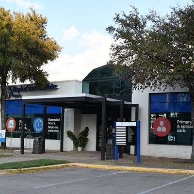
Baylor Scott & White Clinic - Austin North Burnet
2608 Brockton Dr , Austin, TX, 78758- Monday: 8:00 am - 5:00 pm
- Tuesday: 8:00 am - 5:00 pm
- Wednesday: 8:00 am - 5:00 pm
- Thursday: 8:00 am - 5:00 pm
- Friday: 8:00 am - 5:00 pm

Baylor Scott & White Clinic - Austin Oak Hill
5251 US 290 , Austin, TX, 78735- Monday: 8:00 am - 5:00 pm
- Tuesday: 8:00 am - 5:00 pm
- Wednesday: 8:00 am - 5:00 pm
- Thursday: 8:00 am - 5:00 pm
- Friday: 8:00 am - 5:00 pm
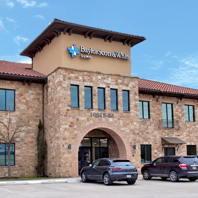
Baylor Scott & White Clinic - Austin River Place
10815 Ranch Rd 2222 , Austin, TX, 78730- Monday: 8:00 am - 5:00 pm
- Tuesday: 8:00 am - 5:00 pm
- Wednesday: 8:00 am - 5:00 pm
- Thursday: 8:00 am - 5:00 pm
- Friday: 8:00 am - 5:00 pm

Baylor Scott & White Clinic - Brenham Hwy 290
604 US 290 , Brenham, TX, 77833- Monday: 7:00 am - 7:00 pm
- Tuesday: 7:00 am - 5:00 pm
- Wednesday: 7:00 am - 5:00 pm
- Thursday: 7:00 am - 7:00 pm
- Friday: 7:00 am - 5:00 pm
- Saturday: 8:00 am - 12:00 pm

Baylor Scott & White Clinic - Buda Medical Center
5330 Overpass Rd Ste 100, Buda, TX, 78610- Monday: 8:00 am - 5:00 pm
- Tuesday: 8:00 am - 5:00 pm
- Wednesday: 8:00 am - 5:00 pm
- Thursday: 8:00 am - 5:00 pm
- Friday: 8:00 am - 5:00 pm
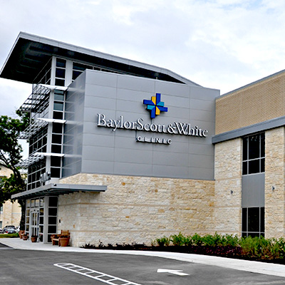
Baylor Scott & White Clinic - Cedar Park
910 E Whitestone Blvd , Cedar Park, TX, 78613- Monday: 8:00 am - 5:00 pm
- Tuesday: 8:00 am - 5:00 pm
- Wednesday: 8:00 am - 5:00 pm
- Thursday: 8:00 am - 5:00 pm
- Friday: 8:00 am - 5:00 pm
- Monday: 7:00 am - 4:30 pm
- Tuesday: 7:00 am - 4:30 pm
- Wednesday: 7:00 am - 4:30 pm
- Thursday: 7:00 am - 4:30 pm
- Friday: 7:00 am - 4:30 pm
- Saturday: 9:00 am - 4:30 pm
- Sunday: 9:00 am - 4:30 pm

Baylor Scott & White Clinic - College Station Rock Prairie
800 Scott and White Dr , College Station, TX, 77845- Monday: 7:30 am - 5:00 pm
- Tuesday: 7:30 am - 5:00 pm
- Wednesday: 7:30 am - 5:00 pm
- Thursday: 7:30 am - 5:00 pm
- Friday: 7:30 am - 5:00 pm

Baylor Scott & White Clinic - Copperas Cove
239 W US Hwy 190 , Copperas Cove, TX, 76522- Monday: 7:45 am - 5:00 pm
- Tuesday: 7:45 am - 5:00 pm
- Wednesday: 7:45 am - 5:00 pm
- Thursday: 7:45 am - 5:00 pm
- Friday: 7:45 am - 5:00 pm
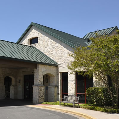
Baylor Scott & White Clinic - Georgetown
4945 Williams Dr , Georgetown, TX, 78633- Monday: 7:30 am - 5:00 pm
- Tuesday: 7:30 am - 5:00 pm
- Wednesday: 7:30 am - 5:00 pm
- Thursday: 7:30 am - 5:00 pm
- Friday: 7:30 am - 5:00 pm

Baylor Scott & White Clinic - Pflugerville Medical Center
2600 E Pflugerville Pkwy Ste 200, Pflugerville, TX, 78660- Monday: 8:00 am - 5:00 pm
- Tuesday: 8:00 am - 5:00 pm
- Wednesday: 8:00 am - 5:00 pm
- Thursday: 8:00 am - 5:00 pm
- Friday: 8:00 am - 5:00 pm
- Monday: 7:30 am - 4:00 pm
- Tuesday: 7:30 am - 4:00 pm
- Wednesday: 7:30 am - 4:00 pm
- Thursday: 7:30 am - 4:00 pm
- Friday: 7:30 am - 4:00 pm
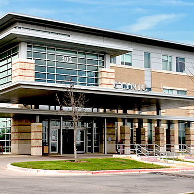
Baylor Scott & White Clinic - Round Rock 302 University
302 University Blvd , Round Rock, TX, 78665- Monday: 8:00 am - 5:00 pm
- Tuesday: 8:00 am - 5:00 pm
- Wednesday: 8:00 am - 5:00 pm
- Thursday: 8:00 am - 5:00 pm
- Friday: 8:00 am - 5:00 pm
Baylor Scott & White Clinic - Taylor
403 Mallard Ln , Taylor, TX, 76574- Monday: 7:30 am - 5:00 pm
- Tuesday: 7:30 am - 5:00 pm
- Wednesday: 7:30 am - 5:00 pm
- Thursday: 7:30 am - 5:00 pm
- Friday: 7:30 am - 4:00 pm
Baylor Scott & White Clinic - Temple
2401 S 31st St , Temple, TX, 76508- Monday: 8:00 am - 5:00 pm
- Tuesday: 8:00 am - 5:00 pm
- Wednesday: 8:00 am - 5:00 pm
- Thursday: 8:00 am - 5:00 pm
- Friday: 8:00 am - 5:00 pm