What is heart valve disease?
Heart valve disease happens when one or more of the valves in your heart aren’t working properly. If not treated, it can cause your heart to work harder, reduce blood flow and even become life-threatening over time.
Your heart has four valves that help keep blood flowing in the right direction. These valves are made of tiny flaps of tissue (called leaflets) that open and close during each heartbeat. The leaflets open to let blood move forward and close to stop blood from flowing backward.
The four valves in your heart are:
- Tricuspid valve: Between the right atrium and right ventricle
- Pulmonary valve: Between the right ventricle and the pulmonary artery
- Mitral valve: Between the left atrium and left ventricle
- Aortic valve: Between the left ventricle and the aorta
Heart valve disease can often be treated if it becomes severe enough to cause symptoms or damage the heart. Your healthcare provider may recommend surgery or a minimally invasive procedure to repair or replace the affected valve. These treatments can restore normal heart function and help you get back to living your life.
Types of heart valve disease
Stenosis (narrowing)
Heart valves can become stiff and narrow, making it harder for blood to flow through. This condition, called valve stenosis, prevents the valve from opening fully and limits blood flow. In severe cases, the valve opening may become so small that the body doesn’t get enough blood.
- Pulmonary valve stenosis: A narrowed pulmonary valve restricts blood flow from the right ventricle to the lungs, making it harder for blood to pick up oxygen and increasing pressure in the heart. This is typically found in people with congenital heart conditions, which are usually present since birth.
- Mitral valve stenosis: A narrowed mitral valve limits blood flow from the left atrium to the left ventricle, leading to fatigue, shortness of breath and fluid buildup in the lungs. This usually occurs when young people have had a history of rheumatic fever or in older people with severe calcification.
- Aortic valve stenosis: A narrowed aortic valve restricts blood flow from the heart to the body, causing the left ventricle to work harder and eventually weaken.
Regurgitation (leaking)
When heart valves don't close properly, blood can flow backward instead of moving forward. This backward flow, called regurgitation, disrupts the heart's one-way blood flow system. It puts extra strain on your heart, making it harder to pump efficiently and reducing the oxygen-rich blood your body needs.
- Tricuspid valve regurgitation: If the tricuspid valve doesn't close fully, blood that's supposed to move from the right ventricle to the lungs leaks backward into the right atrium, causing the atrium to enlarge over time and fluid to build up in the liver, abdomen and legs.
- Pulmonary valve regurgitation: When the pulmonary valve doesn't close properly, blood pumped from the heart to the lungs can leak back into the heart. This mix of oxygen-poor and oxygen-rich blood reduces the amount of oxygen circulating in your body.
- Mitral valve regurgitation: In this condition, some blood leaks backward into the left atrium and then the lungs instead of flowing out to the body, increasing pressure and blood volume in the atrium, potentially causing it to enlarge and leading to fluid buildup in the lungs.
- Aortic valve regurgitation: If the aortic valve doesn't close completely, oxygen-rich blood flows backward into the left ventricle, making the heart work harder to compensate, which, over time, can lead to the left ventricle becoming enlarged, weakened, and increase the risk of heart failure.
Prolapse (slipping out of place)
Prolapse happens when the flaps (leaflets) of a heart valve move out of place or bulge, preventing the valve from closing properly. This can cause blood to flow backward through the valve, disrupting the heart’s normal one-way blood flow.
- Mitral valve prolapse: In mitral valve prolapse, the mitral valve doesn’t close evenly. Instead, part or all of the valve bulges upward into the left atrium when the heart pumps blood. This may allow a small amount of blood to flow backward (called regurgitation). Mitral valve prolapse is also known as click-murmur syndrome, Barlow’s syndrome or floppy valve syndrome. In most cases, mitral valve prolapse has favorable outcomes and rarely progresses into a more serious condition.
- Other valve prolapses: Prolapse of the tricuspid, pulmonary or aortic valves is much less common than mitral valve prolapse. However, they can also cause the valve leaflets to fail to close tightly, leading to a similar issue with blood flow.
Heart valve disease symptoms
Heart valve diseases tend to share symptoms, regardless of which valve is affected. Those symptoms include:
- Angina (chest pain)
- Arrhythmia/Palpitations (irregular heartbeat)
- Dizziness
- Fatigue
- Fainting
- Lightheadedness
- Shortness of breath and other breathing difficulties with exertion or with lying flat on your back
- Swelling in your ankles, belly or feet
For many people, these symptoms develop over time. Some people may not develop symptoms at all.
What causes heart valve disease?
Valve problems can happen for different reasons. Some people are born with them (congenital), while others develop them as they age or because of another health condition.
Some people are born with valve conditions like bicuspid aortic valve disease (two leaflets instead of three) or Marfan syndrome, which affects connective tissue and leads to valve issues. Acquired causes include chest radiation therapy and age-related wear and tear, which can narrow or weaken valves.
Some forms of the disease can develop after you have a medical condition, such as:
- Infective endocarditis, a bacterial infection in the lining of the heart
- Lupus, rheumatoid arthritis, mixed connective tissue disorder and other autoimmune diseases
- Radiation therapy to the chest
- Rheumatic fever, caused by scarring from untreated strep throat
What are the complications of heart valve disease?
Addressing heart valve disease can help protect your health in the long term. When your heart valves don’t function well, your heart experiences additional stress and in rare cases, blood clots. Left untreated, heart valve problems can increase your risk for:
- Heart rhythm abnormalities (atrial fibrillation)
- Congestive heart failure
- Stroke
Diagnosing heart valve disease
Medical history and physical exam
Your provider will listen to your heart to see if you have a heart murmur or an irregular heartbeat. If one or both exist, your provider will want you to have additional diagnostic tests.
Your doctor will talk with you about your symptoms, any heart valve conditions that may run in your family and factors that could increase your risk.
During the physical exam, your doctor may check for:
- Fluid in your lungs: This could indicate your heart isn’t pumping effectively.
- An enlarged heart: This may happen if your heart is working harder than normal.
- A heart murmur: This sound could mean blood is flowing through a valve that is too narrow or not closing properly.
- Swelling in your ankles: This can be a sign of fluid buildup caused by your heart not working as well as it should.
Electrocardiogram
An electrocardiogram, or EKG, is a quick and painless test that measures the electrical activity of your heart. It shows how fast your heart is beating, whether your heartbeat is steady or irregular, and how well the electrical signals are moving through your heart. Healthcare providers often use an EKG during a routine checkup to look for signs of heart problems.
Echocardiogram
An echocardiogram is an imaging test that uses high-frequency sound waves to show doctors how well your heart pumps blood. Sometimes two types of echocardiograms might be used:
- Transthoracic echocardiogram (TTE): The most common type of echocardiogram, this noninvasive procedure is performed outside the body. It's usually a good first test for your provider to evaluate valvular regurgitation and stenosis.
- Transesophageal echocardiogram (TEE): This minimally invasive procedure takes pictures from inside the chest as the ultrasound probe is placed in your esophagus. It's often used when a TTE doesn't provide enough detail, or to assess any prosthetic function after a procedure.
Stress test
A heart stress test shows how well your heart functions when under physical or medication-induced stress. You’ll have electrodes attached to your chest and then be asked to walk on a treadmill or receive a medication that has the same effect on your heart as exercises. Your heart rate, heart rhythm, blood pressure and function of the heart are monitored for signs of heart valve disease.
Cardiac catheterization
Interventional cardiologists perform cardiac catheterization, a minimally invasive procedure, to identify valve defects and other indicators of heart valve disease, such as heart artery blockages and/or pressures inside the various heart chambers.
During cardiac catheterization, a small tube called a catheter is inserted into the body through a blood vessel at the top of your leg or wrist. Your interventional cardiologist guides the catheter to your heart or a blood vessel and performs different procedures to identify problems and treat them effectively.
Imaging tests
Several imaging tests can help diagnose and assess heart valve problems:
- CT scan: Useful in planning surgeries or minimally invasive heart valve procedures to give more detail than an echocardiogram. A CT scan creates detailed images of heart valves to evaluate stenosis, regurgitation, and detect lesions or tumors affecting valve function.
- Chest X-ray: Shows the heart’s size, shape and calcium deposits, which can indicate valve issues like an enlarged or thickened heart.
- Cardiac MRI (CMR): A radiation-free test that uses magnets and radio waves to assess valve disease, measure severity and evaluate how well the heart pumps blood.
What is the treatment for heart valve disease?
Damage to a heart valve cannot be reversed, surgical procedures may be considered to help repair or replace the damaged area.
Your doctor will work with you to identify a care plan and treatment options that best suit your condition, based on your health goals and other underlying health factors that need to be considered.
Medications
Most valve conditions can't be treated with medication alone. However, if the condition isn't severe enough to need surgery but still causes bothersome symptoms or risks, medication may help manage it for a while.
Medications may be prescribed to:
- Ease uncomfortable symptoms of milder valve problems. Diuretics can reduce excess fluid in your body, decreasing the amount of blood the heart has to pump. This lowers your blood pressure and improves your volume status.
- Help maintain a steady heart rhythm if an arrhythmia (irregular heartbeat) is present
- Reduce the risk of blood clots and stroke
Surgery
If your heart valve disease has a significant impact on your quality of life, your cardiologist may recommend having a heart valve repair or replacement surgery or procedure. In addition to open heart surgery, advances in technology and innovation have introduced minimally invasive robotic and transcatheter procedures as options.
Your doctor will walk you through your surgical options, which take the valve or valves needing care, your age, the severity of your heart valve disease and other health factors into consideration.
- Balloon valvuloplasty: During a balloon valvuloplasty, your surgeon uses a narrow, flexible catheter to guide a balloon to your heart valve, where the balloon is expanded to help open the flaps of the heart valve. This procedure is not common for aortic valve stenosis, but may be used if you are living with mitral valve regurgitation.
- Annuloplasty: An annuloplasty can be performed through open-heart surgery or minimally invasive surgery. During the procedure, your surgeon sews a band designed to reshape or reinforce the existing ring around your valve to improve its functionality and correct blood flow. This procedure is not used for aortic valve stenosis.
- Transcatheter aortic valve replacement (TAVR): TAVR is a minimally invasive surgery that allows your heart surgeon to repair a damaged aortic valve. Using a catheter, the surgeon guides a new valve to the site of the damaged valve and expands it, pushing the old valve out of the way.
- Transcatheter edge to edge repair (TEER): This procedure uses a similar approach to TAVR but serves to repair damage to the mitral valve. During the procedure, a catheter is used to guide a device that clips mitral valve leaflets together to improve valve closure and reduce leaking.
Recovering from heart valve disease
You may benefit from cardiac rehab, a medically supervised exercise program designed to help you recover from your procedure. You’ll learn healthy lifestyle habits and stress management techniques with additional guidance on medications to help your heart recover and stay as healthy as possible.
Download our heart valve disease and treatments options guide
Find a location near you
If you have heart valve disease, you have many options for treatment at Baylor Scott & White. Our goal is to restore your heart’s function, help you regain your energy levels and help improve your quality of life.

Baylor Scott & White Advanced Heart Failure Clinic - Abilene
1219 E South 11th St Ste B2, Abilene, TX, 79602
Baylor Scott & White Advanced Heart Failure Clinic - Amarillo
1901 Medi Park Dr Ste 2051, Amarillo, TX, 79106
Baylor Scott & White Advanced Heart Failure Clinic - Dallas
3410 Worth St Ste 250, Dallas, TX, 75246- Monday: 8:00 am - 4:30 pm
- Tuesday: 8:00 am - 4:30 pm
- Wednesday: 8:00 am - 4:30 pm
- Thursday: 8:00 am - 4:30 pm
- Friday: 8:00 am - 4:30 pm

Baylor Scott & White Advanced Heart Failure Clinic - Fort Worth
1400 8th Ave Ste 105, Fort Worth, TX, 76104- Monday: 8:00 am - 4:30 pm
- Tuesday: 8:00 am - 4:30 pm
- Wednesday: 8:00 am - 4:30 pm
- Thursday: 8:00 am - 4:30 pm
- Friday: 8:00 am - 4:30 pm

Baylor Scott & White Advanced Heart Failure Clinic - Longview
906 Judson Rd , Longview, TX, 75601
Baylor Scott & White Advanced Heart Failure Clinic - Lubbock
3711 22nd St Ste B, Lubbock, TX, 79410
Baylor Scott & White Advanced Heart Failure Clinic - Midland/Odessa
420 E 6th St Ste 102, Odessa, TX, 79761
Baylor Scott & White Advanced Heart Failure Clinic - Tyler
1321 S Beckham Ave , Tyler, TX, 75701
Baylor Scott & White Advanced Heart Failure Specialists - Fort Worth
1250 8th Ave Ste 200, Fort Worth, TX, 76104- Monday: 8:00 am - 5:00 pm
- Tuesday: 8:00 am - 5:00 pm
- Wednesday: 8:00 am - 5:00 pm
- Thursday: 8:00 am - 5:00 pm
- Friday: 8:00 am - 5:00 pm

Baylor Scott & White All Saints Medical Center - Fort Worth
1400 8th Ave , Fort Worth, TX, 76104
Baylor Scott & White Arrhythmia Management - Denton
3333 Colorado Blvd , Denton, TX, 76210- Monday: 8:00 am - 5:00 pm
- Tuesday: 8:00 am - 5:00 pm
- Wednesday: 8:00 am - 5:00 pm
- Thursday: 8:00 am - 5:00 pm
- Friday: 8:00 am - 5:00 pm

Baylor Scott & White Arrhythmia Management - Garland
7217 Telecom Pkwy Ste 205, Garland, TX, 75044- Monday: 8:00 am - 5:00 pm
- Tuesday: 8:00 am - 5:00 pm
- Wednesday: 8:00 am - 5:00 pm
- Thursday: 8:00 am - 5:00 pm
- Friday: 8:00 am - 5:00 pm

Baylor Scott & White Arrhythmia Management - Grapevine
2020 W State Hwy 114 Ste 320, Grapevine, TX, 76051
Baylor Scott & White Arrhythmia Management - McKinney
5236 W University Dr POB I, Ste 4900, McKinney, TX, 75071- Monday: 8:00 am - 5:00 pm
- Tuesday: 8:00 am - 5:00 pm
- Wednesday: 8:00 am - 5:00 pm
- Thursday: 8:00 am - 5:00 pm
- Friday: 8:00 am - 5:00 pm

Baylor Scott & White Arrhythmia Management - Plano
1820 Preston Park Blvd Ste 1450, Plano, TX, 75093- Monday: 8:00 am - 5:00 pm
- Tuesday: 8:00 am - 5:00 pm
- Wednesday: 8:00 am - 5:00 pm
- Thursday: 8:00 am - 5:00 pm
- Friday: 8:00 am - 5:00 pm

Baylor Scott & White Arrhythmia Management - Prosper
111 S Preston Rd Ste 10, Prosper, TX, 75078- Monday: 8:00 am - 5:00 pm
- Tuesday: 8:00 am - 5:00 pm
- Wednesday: 8:00 am - 5:00 pm
- Thursday: 8:00 am - 5:00 pm
- Friday: 8:00 am - 5:00 pm

Baylor Scott & White Arrhythmia Management - Rockwall
1005 W Ralph Hall Pkwy Ste 225, Rockwall, TX, 75032
Baylor Scott & White Cardiac Surgery - Dallas
621 N Hall St Ste 120, Dallas, TX, 75226- Monday: 8:30 am - 4:30 pm
- Tuesday: 8:30 am - 4:30 pm
- Wednesday: 8:30 am - 4:30 pm
- Thursday: 8:30 am - 4:30 pm
- Friday: 8:30 am - 4:30 pm

Baylor Scott & White Cardiology Consultants of Texas - Dallas
621 N Hall St Ste 500, Dallas, TX, 75226- Monday: 8:00 am - 5:00 pm
- Tuesday: 8:00 am - 5:00 pm
- Wednesday: 8:00 am - 5:00 pm
- Thursday: 8:00 am - 5:00 pm
- Friday: 8:00 am - 5:00 pm
- Monday: 8:30 am - 4:30 pm
- Tuesday: 8:30 am - 4:30 pm
- Wednesday: 8:30 am - 4:30 pm
- Thursday: 8:30 am - 4:30 pm

Baylor Scott & White Cardiology Consultants of Texas - Greenville
4400 Interstate 30 W Ste 300, Greenville, TX, 75402- Monday: 8:30 am - 5:00 pm
- Tuesday: 8:30 am - 5:00 pm
- Wednesday: 8:30 am - 5:00 pm
- Thursday: 8:30 am - 5:00 pm
- Friday: 8:30 am - 5:00 pm

Baylor Scott & White Cardiology Consultants of Texas - Mesquite
1575 Interstate 30 , Mesquite, TX, 75150- Monday: 8:30 am - 5:00 pm
- Tuesday: 8:30 am - 5:00 pm
- Wednesday: 8:30 am - 5:00 pm
- Thursday: 8:30 am - 5:00 pm
- Friday: 8:30 am - 5:00 pm

Baylor Scott & White Cardiology Consultants of Texas - Midway
4431 E US Hwy 287 , Midlothian, TX, 76065- Monday: 8:00 am - 5:00 pm
- Tuesday: 8:00 am - 5:00 pm
- Wednesday: 8:00 am - 5:00 pm
- Thursday: 8:00 am - 5:00 pm
- Friday: 8:00 am - 5:00 pm

Baylor Scott & White Cardiology Consultants of Texas - Park Cities
9101 N Central Expy Ste 300C, Dallas, TX, 75231- Monday: 8:30 am - 5:00 pm
- Tuesday: 8:30 am - 5:00 pm
- Wednesday: 8:30 am - 5:00 pm
- Thursday: 8:30 am - 5:00 pm
- Friday: 8:30 am - 5:00 pm

Baylor Scott & White Cardiology Consultants of Texas - Red Oak
301 E Ovilla Rd Ste 100, Red Oak, TX, 75154- Monday: 8:00 am - 5:00 pm
- Tuesday: 8:00 am - 5:00 pm
- Wednesday: 8:00 am - 5:00 pm
- Thursday: 8:00 am - 5:00 pm
- Friday: 8:00 am - 5:00 pm
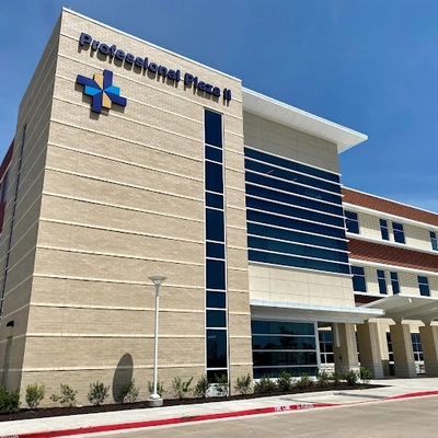
Baylor Scott & White Cardiology Consultants of Texas - Waxahachie
2360 N Interstate 35E Ste 110, Waxahachie, TX, 75165- Monday: 8:00 am - 5:00 pm
- Tuesday: 8:00 am - 5:00 pm
- Wednesday: 8:00 am - 5:00 pm
- Thursday: 8:00 am - 5:00 pm
- Friday: 8:00 am - 5:00 pm

Baylor Scott & White Cardiothoracic Surgery - Irving
1110 Cottonwood Ln Ste 215, Irving, TX, 75038- Monday: 8:30 am - 4:30 pm
- Tuesday: 8:30 am - 4:30 pm
- Wednesday: 8:30 am - 4:30 pm
- Thursday: 8:30 am - 4:30 pm
- Friday: 8:30 am - 4:30 pm

Baylor Scott & White Cardiovascular Associates - Fort Worth
1307 8th Ave Ste 406, Fort Worth, TX, 76104- Monday: 8:00 am - 4:30 pm
- Tuesday: 8:00 am - 4:30 pm
- Wednesday: 8:00 am - 4:30 pm
- Thursday: 8:00 am - 4:30 pm
- Friday: 8:00 am - 4:30 pm

Baylor Scott & White Cardiovascular Consultants - Denton
3333 Colorado Blvd , Denton, TX, 76210- Monday: 8:30 am - 5:00 pm
- Tuesday: 8:30 am - 5:00 pm
- Wednesday: 8:30 am - 5:00 pm
- Thursday: 8:30 am - 5:00 pm
- Friday: 8:30 am - 5:00 pm

Baylor Scott & White Cardiovascular Consultants - Flower Mound
4421 Long Prairie Rd Ste 200, Flower Mound, TX, 75028- Monday: 8:30 am - 5:00 pm
- Tuesday: 8:30 am - 5:00 pm
- Wednesday: 8:30 am - 5:00 pm
- Thursday: 8:30 am - 5:00 pm
- Friday: 8:30 am - 5:00 pm

Baylor Scott & White Cardiovascular Consultants - Grapevine
2020 W State Hwy 114 Ste 200, Grapevine, TX, 76051- Monday: 8:00 am - 5:00 pm
- Tuesday: 8:00 am - 5:00 pm
- Wednesday: 8:00 am - 5:00 pm
- Thursday: 8:00 am - 5:00 pm
- Friday: 8:00 am - 4:00 pm

Baylor Scott & White Cardiovascular Consultants - Keller
620 S Main St Ste 240, Keller, TX, 76248- Monday: 8:00 am - 5:00 pm
- Tuesday: 8:00 am - 5:00 pm
- Wednesday: 8:00 am - 5:00 pm
- Thursday: 8:00 am - 5:00 pm
- Friday: 8:00 am - 4:00 pm

Baylor Scott & White Cardiovascular Consultants - Plano
6000 W Spring Creek Pkwy Ste 220, Plano, TX, 75024- Monday: 8:30 am - 5:00 pm
- Tuesday: 8:30 am - 5:00 pm
- Wednesday: 8:30 am - 5:00 pm
- Thursday: 8:30 am - 5:00 pm
- Friday: 8:30 am - 5:00 pm

Baylor Scott & White Cardiovascular Consultants - Plano II
4716 Alliance Blvd Ste 340, Plano, TX, 75093- Monday: 8:30 am - 4:45 pm
- Tuesday: 8:30 am - 4:45 pm
- Wednesday: 8:30 am - 4:45 pm
- Thursday: 8:30 am - 4:45 pm
- Friday: 8:30 am - 4:45 pm

Baylor Scott & White Cardiovascular Consultants at The Star
3800 Gaylord Pkwy Ste 910, Frisco, TX, 75034- Monday: 8:30 am - 5:00 pm
- Tuesday: 8:30 am - 5:00 pm
- Wednesday: 8:30 am - 5:00 pm
- Thursday: 8:30 am - 5:00 pm
- Friday: 8:30 am - 5:00 pm

Baylor Scott & White Cardiovascular Specialists - Mesquite
5308 N Galloway Ave Ste 201, Mesquite, TX, 75150- Monday: 8:00 am - 5:00 pm
- Tuesday: 8:00 am - 5:00 pm
- Wednesday: 8:00 am - 5:00 pm
- Thursday: 8:00 am - 5:00 pm
- Friday: 8:00 am - 5:00 pm

Baylor Scott & White Cardiovascular Specialists - Rockwall
6705 Heritage Pkwy Ste 202, Rockwall, TX, 75087- Monday: 8:00 am - 5:00 pm
- Tuesday: 8:00 am - 5:00 pm
- Wednesday: 8:00 am - 5:00 pm
- Thursday: 8:00 am - 5:00 pm
- Friday: 8:00 am - 5:00 pm

Baylor Scott & White Clinic - Austin Downtown
200 E Cesar Chavez St Ste G140, Austin, TX, 78701- Monday: 8:00 am - 5:00 pm
- Tuesday: 8:00 am - 5:00 pm
- Wednesday: 8:00 am - 5:00 pm
- Thursday: 8:00 am - 5:00 pm
- Friday: 8:00 am - 5:00 pm

Baylor Scott & White Clinic - Austin North Burnet
2608 Brockton Dr , Austin, TX, 78758- Monday: 8:00 am - 5:00 pm
- Tuesday: 8:00 am - 5:00 pm
- Wednesday: 8:00 am - 5:00 pm
- Thursday: 8:00 am - 5:00 pm
- Friday: 8:00 am - 5:00 pm

Baylor Scott & White Clinic - Austin Oak Hill
5251 US 290 , Austin, TX, 78735- Monday: 8:00 am - 5:00 pm
- Tuesday: 8:00 am - 5:00 pm
- Wednesday: 8:00 am - 5:00 pm
- Thursday: 8:00 am - 5:00 pm
- Friday: 8:00 am - 5:00 pm

Baylor Scott & White Clinic - Austin River Place
10815 Ranch Rd 2222 , Austin, TX, 78730- Monday: 8:00 am - 5:00 pm
- Tuesday: 8:00 am - 5:00 pm
- Wednesday: 8:00 am - 5:00 pm
- Thursday: 8:00 am - 5:00 pm
- Friday: 8:00 am - 5:00 pm

Baylor Scott & White Clinic - Brenham Hwy 290
604 US 290 , Brenham, TX, 77833- Monday: 7:00 am - 7:00 pm
- Tuesday: 7:00 am - 5:00 pm
- Wednesday: 7:00 am - 5:00 pm
- Thursday: 7:00 am - 7:00 pm
- Friday: 7:00 am - 5:00 pm
- Saturday: 8:00 am - 12:00 pm

Baylor Scott & White Clinic - Buda Medical Center
5330 Overpass Rd Ste 100, Buda, TX, 78610- Monday: 8:00 am - 5:00 pm
- Tuesday: 8:00 am - 5:00 pm
- Wednesday: 8:00 am - 5:00 pm
- Thursday: 8:00 am - 5:00 pm
- Friday: 8:00 am - 5:00 pm

Baylor Scott & White Clinic - Cedar Park
910 E Whitestone Blvd , Cedar Park, TX, 78613- Monday: 8:00 am - 5:00 pm
- Tuesday: 8:00 am - 5:00 pm
- Wednesday: 8:00 am - 5:00 pm
- Thursday: 8:00 am - 5:00 pm
- Friday: 8:00 am - 5:00 pm
- Monday: 7:00 am - 4:30 pm
- Tuesday: 7:00 am - 4:30 pm
- Wednesday: 7:00 am - 4:30 pm
- Thursday: 7:00 am - 4:30 pm
- Friday: 7:00 am - 4:30 pm
- Saturday: 9:00 am - 4:30 pm
- Sunday: 9:00 am - 4:30 pm

Baylor Scott & White Clinic - College Station Rock Prairie
800 Scott and White Dr , College Station, TX, 77845- Monday: 7:30 am - 5:00 pm
- Tuesday: 7:30 am - 5:00 pm
- Wednesday: 7:30 am - 5:00 pm
- Thursday: 7:30 am - 5:00 pm
- Friday: 7:30 am - 5:00 pm

Baylor Scott & White Clinic - Copperas Cove
239 W US Hwy 190 , Copperas Cove, TX, 76522- Monday: 7:45 am - 5:00 pm
- Tuesday: 7:45 am - 5:00 pm
- Wednesday: 7:45 am - 5:00 pm
- Thursday: 7:45 am - 5:00 pm
- Friday: 7:45 am - 5:00 pm

Baylor Scott & White Clinic - Georgetown
4945 Williams Dr , Georgetown, TX, 78633- Monday: 7:30 am - 5:00 pm
- Tuesday: 7:30 am - 5:00 pm
- Wednesday: 7:30 am - 5:00 pm
- Thursday: 7:30 am - 5:00 pm
- Friday: 7:30 am - 5:00 pm

Baylor Scott & White Clinic - Pflugerville Medical Center
2600 E Pflugerville Pkwy Ste 200, Pflugerville, TX, 78660- Monday: 8:00 am - 5:00 pm
- Tuesday: 8:00 am - 5:00 pm
- Wednesday: 8:00 am - 5:00 pm
- Thursday: 8:00 am - 5:00 pm
- Friday: 8:00 am - 5:00 pm
- Monday: 7:30 am - 4:00 pm
- Tuesday: 7:30 am - 4:00 pm
- Wednesday: 7:30 am - 4:00 pm
- Thursday: 7:30 am - 4:00 pm
- Friday: 7:30 am - 4:00 pm
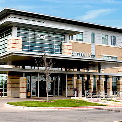
Baylor Scott & White Clinic - Round Rock 302 University
302 University Blvd , Round Rock, TX, 78665- Monday: 8:00 am - 5:00 pm
- Tuesday: 8:00 am - 5:00 pm
- Wednesday: 8:00 am - 5:00 pm
- Thursday: 8:00 am - 5:00 pm
- Friday: 8:00 am - 5:00 pm

Baylor Scott & White Clinic - Taylor
403 Mallard Ln , Taylor, TX, 76574- Monday: 7:30 am - 5:00 pm
- Tuesday: 7:30 am - 5:00 pm
- Wednesday: 7:30 am - 5:00 pm
- Thursday: 7:30 am - 5:00 pm
- Friday: 7:30 am - 4:00 pm

Baylor Scott & White Clinic - Temple
2401 S 31st St , Temple, TX, 76508- Monday: 8:00 am - 5:00 pm
- Tuesday: 8:00 am - 5:00 pm
- Wednesday: 8:00 am - 5:00 pm
- Thursday: 8:00 am - 5:00 pm
- Friday: 8:00 am - 5:00 pm